
Espectro electromagnético
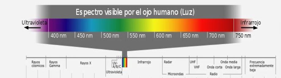
Se llama así al conjunto de todos los tipos de luz, o radiaciones electromagnéticas que existen, las cuales se puede dividir en grandes grupos: radio, microondas, infrarrojo, visible, ultravioleta, rayos X y rayos gamma.

Ilustración 1 Espectro electromagnético
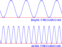
Se diferencian unas de otras por su frecuencia (ver ilustración 2), que es como “la huella dactilar” que identifica a una onda
También se utiliza la longitud de onda (ver ilustración 3), que es la distancia entre dos picos de la misma amplitud y es inversamente proporcional a la frecuencia. Cada longitud de onda es un color, aunque la mayor parte de ellos son invisibles a nuestros ojos. Todos los cuerpos emiten radiación electromagnética cuya frecuencia depende de la temperatura a la que se encuentren: a mayor energía mayor frecuencia.

Las ondas infrarrojas están entre el intervalo de 0,7 a 100 micrómetros. Esta radiación se asocia generalmente con el calor.
Son usadas para algunos sistemas especiales de comunicaciones, como en astronomía para detectar estrellas y otros cuerpos, para guías en armas, en los que se usan detectores de calor para descubrir cuerpos móviles en la oscuridad. También se usan en los controles remotos de los televisores.
Espectro visible es el espectro de radiación electromagnética que es visible para el ojo humano. Cuando estamos viendo un objeto, es porque ese objeto está siendo iluminado por la luz visible. El intervalo visible se considera de los 380 a los 750 nm.
Ultravioleta La luz ultravioleta cubre el intervalo de 4 a 400 nanómetros. El Sol es una importante fuente emisora de esta radiación, la cual causa cáncer de piel en exposiciones prolongadas. Este tipo de onda se usa en aplicaciones del campo de la medicina.
Rayos X Radiación electromagnética, invisible, capaz de atravesar cuerpos opacos. La longitud de onda está entre 10 a 0,1 nanómetros.
Rayos gamma
Radiación electromagnética producida generalmente por elementos radioactivos. Este tipo de radiación se produce también en fenómenos astrofísicos de gran violencia.
Debido a las altas energías que poseen, los rayos gamma constituyen un tipo de radiación ionizante capaz de penetrar más profundamente. Dada su alta energía pueden causar grave daño al núcleo de las células, por lo que son usados para esterilizar equipos médicos y alimentos.
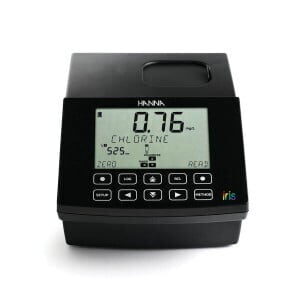
Estas radiaciones se miden especialmente mediante espectrofotómetros, los cuales pueden medir una o más de estas en un solo equipo, por ejemplo el IRIS HI 801 mide principalmente el intervalo de luz visible de 340 a 900 nm, sin embargo hay espectrofotómetros que miden en el intervalo de luz ultra violeta y luz visible, comúnmente llamados UV- VIS y también existen los espectrofotómetros que miden en la radiación infrarroja.
La base de la espectroscopia Visible y Ultravioleta consiste en medir la intensidad del color (o de la radiación absorbida en UV) a una longitud de onda específica comparándola con otras soluciones de concentración conocida (soluciones estándar) que contengan la misma especie absorbente. Para tener esta relación se emplea la Ley de Beer, que establece que para una misma especie absorbente en una celda de espesor constante, la absorbancia es directamente proporcional a la concentración.
La coloración de la solución se debe a la especie absorbente y esta coloración puede ser natural o inducida. La coloración natural puede ser la base de la cuantificación de una especie, como por ejemplo: la clorofila en ciertas plantas, los complejos metálicos que se encuentran presentes en solución acuosa, como son los iones de Cobre (II), Manganeso (VII), Cobalto (III), etc.
Por esta razón es importante tener ciertos conocimientos de la muestra que se desea medir, principalmente si existe alguna metodología certificada con la que se pueda realizar la cuantificación y que sobre todo la muestra a analizar absorba en el intervalo de longitud de onda con que cuenta nuestro espectrofotómetro.
En Hanna contamos con un espectrofotómetro que trabaja en el intervalo de 340 a 900 nm es decir, el intervalo de luz visible y una parte pequeña de los intervalos Ultravioleta e infrarrojo, a continuación se presenta una tabla con más de las especificaciones que presenta el equipo.
Especificaciones
| Intervalo de longitud de onda | 340-900 nm |
| Resolución de longitud de onda | 1 nm |
| Exactitud de longitud de onda | ± 1.5 nm |
| Intervalo fotométrico | 0.000 – 3.000 Abs |
| Exactitud fotométrica | 5 mAbs en 0.000-0.500 Abs 1% en 0.500- 3.000 Abs |
| Modo de medición | Transmitancia (%), Absorbancia, Concentración |
| Celdas para muestra | 10 mm cuadrada, 50 mm rectangular, 16 mm redonda, 22 mm redonda, 13 mm redonda (vial) |
| Selección de la longitud de onda | Automática, basada en el método seleccionado (editable únicamente para métodos del usuario) |
| Fuente de luz | Lámpara halógena de tungsteno |
| Sistema óptico | Haz dividido |
| Calibración de longitud de onda | Interna, automática al encenderse con retroalimentación visual. |
| Luz dispersa | <0.1 %T en 340 nm con NaNO2 |
| Ancho de banda espectral | 5 nm |
| Número de métodos | Hasta 150 de fábrica (85 programados), Hasta 100 de usuario |
| Datos almacenados | 9999 valores medidos |
| Capacidad de exportación | Archivo con formato csv, Archivo con formato pdf |
| Conectividad | 1x USB A (host de almacenamiento masivo), 1x USB B (dispositivo de almacenamiento masivo) |
| Duración de la batería | 3000 mediciones o 8 horas |
| Alimentación eléctrica | Adaptador de corriente de 15 VCD, Batería recargable de ion litio 10.8 VCD |
| Condiciones ambientales | 0 a 50°C (32 a 122°F); 0 a 95% HR |
| Dimensiones | 155 x 205 x 322 mm (6.1 x 8.0 x 12.6″) |
| Peso | 3 kg (6.6 Ibs.) |
Electromagnetic spectrum
This is called the set of all types of light, or electromagnetic radiation that exist, which can be divided into large groups: radio, microwave, infrared, visible, ultraviolet, X-rays and gamma rays (Illustration 1).
Illustration 1 Electromagnetic spectrum
They differ from each other in their frequency (illustration 2), which is like “the fingerprint” that identifies a wave
The wavelength is also used (llustration 3), which is the distance between two peaks of the same amplitude and is inversely proportional to the frequency. Each wavelength is a color, although most of them are invisible to our eyes. All bodies emit electromagnetic radiation whose frequency depends on the temperature at which they are located: the higher the energy, the greater the frequency.

The infrared waves are between the range of 0.7 to 100 micrometers. This radiation is usually associated with heat.
They are used for some special communication systems, such as astronomy to detect stars and other bodies, for guides in weapons, in which heat detectors are used to discover moving bodies in the dark. They are also used in TV remote controls.
Visible spectrum is the spectrum of electromagnetic radiation that is visible to the human eye. When we are seeing an object, it is because that object is being illuminated by visible light. The visible range is considered from 380 to 750 nm (llustration 4).
Ultraviolet light covers the range of 4 to 400 nanometers. The Sun is an important source of this radiation, which causes skin cancer in prolonged exposures. This type of wave is used in applications in the field of medicine.
X rays Electromagnetic radiation, invisible, capable of crossing opaque bodies. The wavelength is between 10 to 0.1 nanometers.
Gamma rays Electromagnetic radiation generally produced by radioactive elements. This type of radiation also occurs in astrophysical phenomena of great violence.
Due to the high energies they possess, gamma rays constitute a type of ionizing radiation capable of penetrate more deeply. Given their high energy can cause serious damage to the nucleus of cells, so they are used to sterilize medical equipment and food.
These radiations are measured especially by spectrophotometers, which can measure one or more of these in a single equipment, for example the IRIS HI 801 mainly measures the visible light range of 340 to 900 nm, however there are spectrophotometers that measure in the interval of ultra violet light and visible light, commonly called UV-VIS and there are also spectrophotometers that measure infrared radiation.
The basis of the Visible and Ultraviolet spectroscopy is to measure the intensity of the color (or of the radiation absorbed in UV) at a specific wavelength compared to other solutions of known concentration (standard solutions) that contain the same absorbing species. To have this relationship, Beer’s Law is used, which establishes that for the same absorbent species in a cell of constant thickness, the absorbance is directly proportional to the concentration.
The coloration of the solution is due to the absorbent species and this coloration can be natural or induced. Natural coloration can be the basis of the quantification of a species, such as: chlorophyll in certain plants, metal complexes that are present in aqueous solution, such as Copper (II), Manganese (VII) ions, Cobalt (III), etc.
For this reason, it is important to have certain knowledge of the sample to be measured, especially if there is a certified methodology with which quantification can be performed and that, above all, the sample to be analyzed absorbs in the wavelength range of our sample. spectrophotometer
In Hanna we have a spectrophotometer that works in the range of 340 to 900 nm that is the visible light interval and a small part of the Ultraviolet and infrared intervals. Below is a table with more specifications of the iris HI801 spectrophotometer.
Specifications
| Wavelength Range | 340 to 900 nm |
| Wavelength Resolution | 1 nm |
| Wavelength Accuracy | ±1.5 nm |
| Measurement Modes | Transmittance (% T), absorbance (abs), concentration with choice of units (ppm, mg/L, ppt, ºf, ºe, ppb, meq/L, μg/L, PCU, Pfund, pH, dKH, ºdH, meq/kg or no measurement unit) |
| Wavelength Selection | automatic, based on the selected method (editable for user methods only) |
| Light Source | tungsten halogen lamp |
| Optical System | split beam sample and reference light detectors |
| Wavelength Calibration | internal, automatic at power-on, visual feedback |
| Stray Light | <0.1 % T at 340 nm with NaNO2 |
| Spectral bandwidth | 5 nm (full width at half maximum) |
| Sample Cell | 16 mm round, 22 mm round, 13 mm vial, 10 mm square, 50 mm rectangular (with automatic detection) |
| Programs (Factory/User) | up to 150 factory (85 pre-loaded); up to 100 user developed |
| Data Points Stored | up to 9999 measured values |
| Export Capability | .csv file format, .pdf file format |
| Connectivity | (1) USB – A (mass storage host); (1) USB – B (mass storage device) |
| Battery Type / Life | 3000 measurements or 8 hours |
| Power Supply | 15 VDC power adapter; 10.8 VDC Li-Ion rechargeable battery |
| Environment | 0 to 50 °C (32 to 122 °F); 0 to 95% RH |
| Dimensions | 155 x 205 x 322 mm (6.1 x 8.0 x 12.6″) |
| Weight | 3 kg (6.6 lbs) |